Every year June 25 is celebrated as the World Vitiligo Day to raise awareness regarding Vitiligo, fight prejudice, and raise funds for research, support & education. The aim of this day is to include the recognition of the bullying, social neglect, psychological trauma, and disability of millions of people affected by vitiligo. The primary purpose of this day is to raise money for research, give free skin exams, and educate physicians on how to best take care of patients with vitiligo.
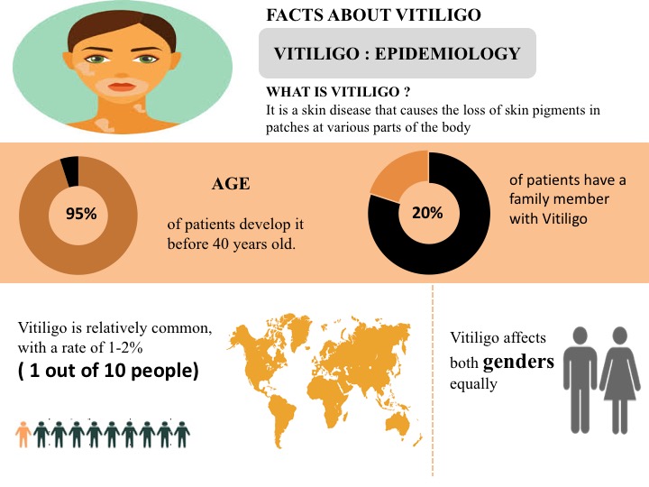
Vitiligo is the long term skin condition in which the skin loses the pigment i.e. melanin, essential for determining the color of skin, hair, and eyes. This leads to the slow growth of the white patches of irregular shapes on the skin. Vitiligo can also affect the mucous membranes, including tissues inside the mouth and nose. It can affect people of all skin types but is generally noticeable much in darker skin people.
Vitiligo Facts
Around 1 out of 10 people i.e. 1-2% of people are suffering from Vitiligo worldwide
The United States accounts for the highest prevalent cases of Vitiligo in comparison to the EU5 (Germany, France, Spain, Italy, and the United Kingdom) and Japan
Vitiligo is classified as the non-segmental, and segmental Vitiligo. Non-segmental Vitiligo is most prevalent in comparison to the segmental Vitiligo
Approximately, 95% of the people develop the condition prior to age 40.
Around 20% of people have a family member suffering from Vitiligo
Males and females are almost equally affected by Vitiligo
Vitiligo is sometimes associated with certain other medical conditions, including thyroid dysfunction, diabetes mellitus, Addison’s disease, etc.
Several factors such as autoimmune disease, genetic factors, sunburn or any cut, oxidative stress, neurochemicals, and exposure to the industrial chemicals increase the risk of developing Vitiligo
June is the Scoliosis Awareness Month that aims at highlighting the growing need for education, early detection, and awareness regarding scoliosis and its prevalence within the community. It unites scoliosis patients, families, physicians, clinicians, institutions, and related businesses in collaborative partnerships of local activities, events, and grassroots networking throughout the month.
Scoliosis Overview
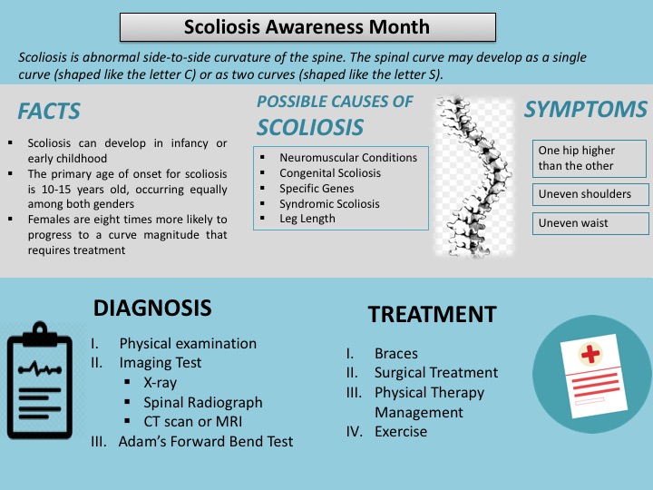
Scoliosis is an abnormal side-to-side curvature of the spine. The spinal curve may develop as a single curve (shaped like the letter C) or as two curves (shaped like the letter S). It is often defined as spinal curvature in the “coronal” (frontal) plane. While the degree of curvature is measured on the coronal plane, scoliosis is actually a more complex, three-dimensional problem which involves the following planes:
Coronal plane
Sagittal plane
Axial plane
The coronal plane is a vertical plane from head to foot and parallel to the shoulders, dividing the body into anterior (front) and posterior (back) sections. The sagittal plane divides the body into right and left halves. The axial plane is parallel to the plane of the ground and at right angles to the coronal and sagittal planes.
Scoliosis is hereditary among the people with scoliosis who are more likely to have children with scoliosis; however, there is no correlation between the severity of the curves from one generation to the next. In children and teens, scoliosis often does not have any noticeable symptoms and may not be noticeable until it has progressed significantly. Most cases of scoliosis are mild, but some spine deformities continue to get more severe as children grow. Severe scoliosis can be disabling. An especially severe spinal curve can reduce the amount of space within the chest, making it difficult for the lungs to function properly. The most common form of scoliosis appears in adolescents. It is known as adolescent idiopathic scoliosis. It can affect children from the age of 10 years.
The symptoms can include the head is slightly off center, the ribcage is not symmetrical, so the ribs may be at different heights and one hip is more prominent than the other. Furthermore, in infants, symptoms can include: a bulge on one side of the chest, consistently lying curved to one side (in babies), Problems with the heart and lungs, leading to shortness of breath and chest pain.
According to the American Association of Neurological Surgeons (AANS), about 80 percent of scoliosis cases have no identifiable cause. The condition is often diagnosed during the first seven years of a child’s life.
Etiology
Neuromuscular Conditions: These affect the nerves and muscles and include cerebral palsy, poliomyelitis, and muscular dystrophy.
Congenital Scoliosis (present at birth): This is rare and occurs because the bones in the spine developed abnormally when the fetus was growing inside the mother.
Specific genes: At least one gene is thought to be involved in scoliosis.
Leg length: If one leg is longer than the other, the individual may develop scoliosis.
Syndromic scoliosis: Scoliosis can develop as part of another disease, including neurofibromatosis and Marfan’s syndrome.
Osteoporosis: This can cause secondary scoliosis due to bone degeneration.
Risk Factors
There are certain risk factors associated with scoliosis include: age, gender & genetics etc. are explained below:
Age: Signs and symptoms often start during a growth spurt just before puberty.
Gender: Females have a higher risk in comparison to the males
Genetics: People with scoliosis may have a close relative with the condition.
Diagnosis
Scoliosis is confirmed through a physical examination, an x-ray, spinal radiograph, CT scan or MRI. The curve is measured by the Cobb Method and is diagnosed in terms of severity by the number of degrees. A positive diagnosis of scoliosis is made based on a coronal curvature measured on a posterior-anterior radiograph of greater than 10 degrees. In general, a curve is considered significant if it is greater than 25 to 30 degrees. Curves exceeding 45 to 50 degrees are considered severe and often require more aggressive treatment.
A standard exam that is sometimes used by pediatricians and in grade school screenings is called the Adam’s Forward Bend Test. During this test, the patient leans forward with his or her feet together and bends 90 degrees at the waist. This is a simple initial screening test that can detect potential problems, but cannot determine accurately the exact type or severity of the deformity. The tests are required for an accurate and positive diagnosis.
Physical Examination
Doctor would check the spine curvature and whether the shoulders and waist area are symmetrical or not.
Imaging Tests
Imaging tests doctor may order to look for scoliosis include:
X-ray: During this test, small amounts of radiation are used to create a picture of your spine.
MRI scan: This test uses radio and magnetic waves to get a detailed picture of bones and the tissue surrounding them.
CT scan: During this test, X-rays are taken at a variety of angles to get a 3-D picture of the body.
Bone scan: This test detects a radioactive solution injected into your blood that concentrates in areas of increased circulation, highlighting spinal abnormalities.
Treatment
Treatment of scoliosis is based on the severity of the curve and the chances of the curve getting worse. Certain types of scoliosis have a greater chance of getting worse, so the type of scoliosis also helps to determine the proper treatment. There are three main categories of treatment i.e. observation, bracing (for example, thoracolumbosacral orthosis or TLSO back brace), and surgery. Consequently, there are treatments available that do not involve surgery, but in some individuals, surgery may be their best option.
Observation
In many children with scoliosis, the spinal curve is mild enough to not require treatment. However, if the doctor is worried that the curve may be increasing, he or she may wish to examine the child every four to six months throughout adolescence.
In adults with scoliosis, X-rays are usually recommended once every five years, unless symptoms are getting progressively worse.
Bracing
Braces are only effective in patients who have not reached skeletal maturity. If the child is still growing and his or her curve is between 25 degrees and 40 degrees, a brace may be recommended to prevent the curve from progressing. There have been improvements in brace design and the newer models fit under the arm, not around the neck. There are several different types of braces available. For optimal effectiveness, the brace should be checked regularly to assure a proper fit and may need to be worn 16 to 23 hours every day until growth stops.
Surgery
In children, the two primary goals of surgery are to stop the curve from progressing during adulthood and to diminish spinal deformity. Most experts would recommend surgery only when the spinal curve is greater than 40 degrees and there are signs of progression. This surgery can be done using an anterior approach (through the front) or a posterior approach (through the back) depending on the particular case.
A number of factors can lead to increased surgical-related risks in older adults with degenerative scoliosis. These factors include the following: advanced age, being a smoker, being overweight and the presence of other health/medical problems. In general, both surgery and recovery time are expected to be longer in older adults with scoliosis.
Following surgical procedures are used for the treatment of scoliosis
Posterior approach: The most frequently performed surgery for adolescent idiopathic scoliosis involves posterior spinal fusion with instrumentation and bone grafting. This is performed through the back while the patient lies on his or her stomach.
Anterior approach: The patient lies on his or her side during the surgery. The surgeon makes incisions in the patient’s side, deflates the lung and removes a rib in order to reach the spine. Video-assisted thoracoscopic (VAT) surgery offers enhanced visualization of the spine and is a less invasive surgery than an open procedure. The anterior spinal approach has several potential advantages: better deformity correction, quicker patient rehabilitation, improved spine mobilization and fusion of fewer segments.
Decompressive laminectomy: The laminae (roof) of the vertebrae are removed to create more space for the nerves. A spinal fusion with or without spinal instrumentation is often recommended when scoliosis and spinal stenosis are present. Various devices (like screws or rods) may be used to enhance fusion and support unstable areas of the spine.
Minimally invasive surgery (MIS) : Fusion can sometimes be performed via smaller incisions through MIS. The use of advanced fluoroscopy (X-ray imaging during surgery) and endoscopy (camera technology) has improved the accuracy of incisions and hardware placement, minimizing tissue trauma while enabling a MIS approach. It is important to keep in mind that not all cases can be treated in this manner and a number of factors contribute to the surgical method used.
The benefits of surgery should always be weighed carefully against its risks. Although a large percentage of scoliosis patients benefit from surgery, there is no guarantee that surgery will stop curve progression and symptoms in every individual.
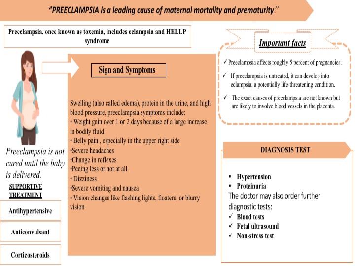
Pre-eclampsia is a leading cause of maternal mortality and prematurity. It is formerly called toxemia, which is when a pregnant woman has high blood pressure, protein in her urine, and swelling in her legs, feet, and hands. It can range from mild to severe. It usually happens late in the pregnancy after 20 weeks of pregnancy, though it can come earlier or just after delivery. In addition, there are several complications associated with preeclampsia such as HELLP syndrome, poor blood flow to the placenta & placental abruption, etc. Preeclampsia can lead to eclampsia, a serious condition that can have health risks for mom and baby and, in rare cases, cause death. Women with preeclampsia who have seizures have eclampsia. Approximately around 5% of all pregnant women get affected by preeclampsia.
The symptoms often begin after 34 weeks. Symptoms develop after birth, usually within 48 hours of delivery in few cases. They tend to go away on their own but can last up to 12 weeks after birth. In addition, the common symptoms develop such as edema, protein in the urine, and high blood pressure, preeclampsia symptoms include weight gain, belly pain, severe headaches, changes in reflexes, peeing less or not at all, dizziness, severe vomiting and nausea, vision changes like flashing lights, floaters, or blurry vision
Yet, the exact cause of preeclampsia is unidentified. But most of them say this problem happens with the development of the placenta because the blood vessels that supply it are narrower than normal and respond differently to hormonal signals. Because the blood vessels are narrower than normal, blood flow is limited. Several factors such as the history of preeclampsia, age, race, chronic hypertension, first pregnancy, obesity, and multiple pregnancies increase the chances of developing the preeclampsia
Preeclampsia is diagnosed through the following tests:
Hypertension: The woman’s blood pressure is raised above 140/90 millimeters of mercury is abnormal in pregnancy.
Proteinuria: Protein is detected in the urine is indicated for the severity of the condition.
The doctor may also recommend some further diagnostic tests:
Blood tests – to assess the kidney and liver functions.
Fetal ultrasound – to monitor the baby’s progress.
Non-stress test – the doctor checks how the baby’s heartbeat reacts when they move. If the heartbeat increases 15 beats or more a minute for at least 15 seconds twice every 20 minutes, it is an indication that everything is normal
Recently, UniSA biomedical engineer Professor Benjamin Thierry developed a range of solid-state sensing and wearable technologies capable of diagnosing conditions including preeclampsia, epilepsy, fetal arrhythmias, and heart attacks. These wearables use a cutting-edge solid-state sensing technology called Field-Effect Transistors, which can measure bioelectric signals with extreme sensitivity when implemented at the nanoscale.”
However, Preeclampsia is not cured until the baby is delivered. Hence, the mother’s blood pressure comes down, she is at a greater risk of stroke, severe bleeding, separation of the placenta separates from the uterus and seizures. In some cases, especially if the preeclampsia started early, the delivery may not be the best option for the fetus.
Supportive therapy is recommended for the women Preeclampsia. Supportive therapy is as follows:
Antihypertensives: These are used to lower blood pressure.
Anticonvulsants: In severe cases, these drugs are used to prevent a first seizure. The doctor may prescribe magnesium sulfate.
Corticosteroids: If the mother has preeclampsia or HELLP syndrome (see below) these drugs can improve platelet and liver functioning. This can prolong pregnancy.
While preeclampsia cannot be fully prevented, there are significant steps a woman can follow to avoid the moderate factors contribute to high blood pressure.
These may include:
drinking between 6 and 8 glasses of water every day
avoiding fried or processed food
excluding added salt from the diet
regular exercise
avoiding alcohol and caffeine intake
keeping the feet elevated a few times per day
resting
supplements and medications as prescribed by your doctor
May, 10-16 is the Food Allergy Awareness Week created by the Food Allergy Research and Education (FARE) to raise awareness regarding the different food allergies and improve public understanding of what can sometimes be a life-threatening condition.
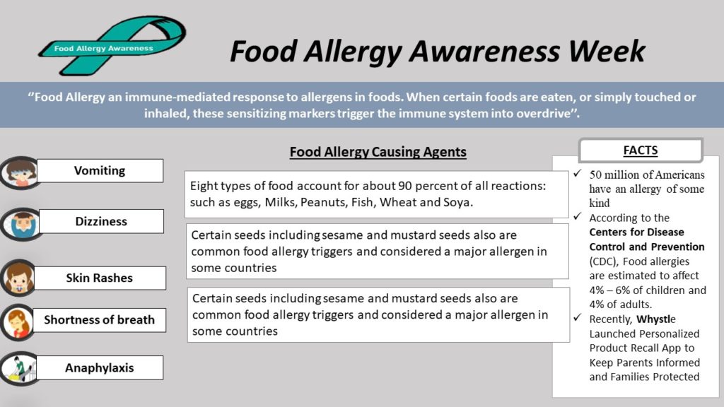
The food allergy is an immune-mediated response to allergens in foods. When certain foods are eaten, or simply touched or inhaled, these sensitizing markers trigger the immune system into overdrive. The prevalence of food allergy is increasing worldwide. Likewise, more than 50 million Americans have an allergy of some kind. Food allergy symptoms are most common in babies and children, but they can appear at any age. It can develop an allergy to foods even eaten for years with no problems.
According to the Centers for Disease Control and Prevention (CDC), Food allergies are estimated to affect 4% – 6% of children and 4% of adults, which occurs when the immune system identifies a food or substance as a danger and triggers a protective response. This reaction may cause a wide range of symptoms, including Vomiting or stomach cramps, hives, shortness of breath, wheezing, repetitive cough, dizziness or feeling faint, weak pulse and anaphylaxis, etc. While any food can cause an adverse reaction, eight types of food account for about 90 percent of all reactions: such as eggs, milk, peanuts, fish, wheat, and soy. In addition, certain seeds including sesame and mustard seeds (the main ingredient in the condiment mustard), also are common food allergy triggers and considered a major allergen in some countries. Food allergy may also involve in affecting some other areas such as skin, the gastrointestinal tract, the cardiovascular system, and the respiratory tract.
Moreover, the food-related symptoms may occur within two hours of ingestion; often they start within minutes. In some very rare cases, the reaction may be delayed by four to six hours or even longer. The delayed reactions are most typically seen in children who develop eczema as a symptom of food allergy and in people with a rare allergy to red meat caused by the bite of a lone star tick.
Another type of delayed food allergy reaction stems from food protein-induced enterocolitis syndrome (FPIES), a severe gastrointestinal reaction that generally occurs two to six hours after consuming milk, soy, certain grains, and some other solid foods. It mostly occurs in young infants who are being exposed to these foods for the first time or who are being weaned.
Sometimes, the diagnosis of FPIES may be delayed. FPIES is a medical emergency that should be treated with IV rehydration. Allergists ask detailed questions about medical history and the symptoms for the diagnosis of food allergy. Various skin tests and/or blood tests are used for the detecting the food-specific immunoglobulin E (IgE) antibodies present in the body i.e. Skin- prick Test and Blood Test
Skin-Prick Tests provide results within 20 minutes. A liquid containing a tiny amount of the food allergen is placed on the skin of the arm or back and then the skin is pricked with a small, sterile probe, allowing the liquid to seep under the skin. The test, which isn’t painful but can be uncomfortable, is considered positive if a wheal develops at the site where the suspected allergen was placed. As a control, they ’ll also get a skin prick with a liquid that doesn’t contain the allergen; this should not provoke a reaction, allowing comparison between the two test sites.
Blood Tests which are less accurate than skin tests, measure the amount of IgE antibody to the specific food being tested. Results are typically come out in about a week and are reported in the numerical value.
The Food Allergy can be managed in the following ways:
To avoid consuming the food that causes problems. In addition, carefully check ingredient labels of food products.
Despite this, there are some other treatments to reduce minor and severe allergic reactions. e.g. For a minor allergic reaction, over-the-counter or prescribed antihistamines to reduce the symptoms. However, it can’t be used to treat severe allergic reactions.
For a severe allergic reaction, recommended an emergency injection of epinephrine and a trip to the emergency room. In addition, many people with allergies carry an epinephrine autoinjector (Adrenaclick, EpiPen)
During this Food Allergy Awareness Week, Whystle Launched Personalized Product Recall App to Keep Parents Informed and Families Protected. This app provides personalized safety information and up-to-the-minute recall notices, especially for up-to-date allergen recalls. Besides, every three minutes, food allergy reactions send someone to the emergency room.
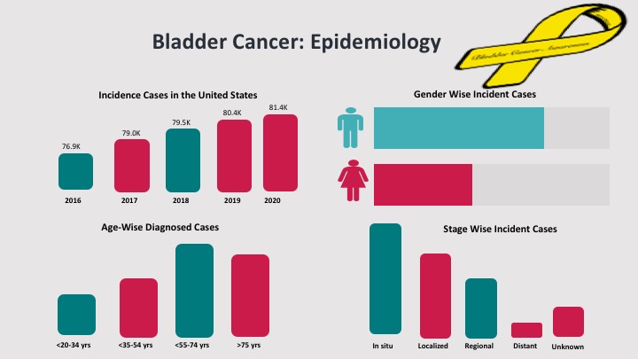
May is the Bladder Cancer Awareness month to raise awareness regarding types, etiology, risk factors, diagnosis, treatment, and preventative measures for Bladder Cancer.
Bladder Cancer is the most common cancer which includes the uncontrollable cell growth in the urinary bladder and spreads to the other parts of the body. According to the Surveillance, Epidemiology, and End Results (SEER), the incidence cases of Bladder Cancer is expected to be 81,400 in 2020. Around 17,980 people are expected to die due to Bladder Cancer in 2020.
According to the Bladder Cancer Advocacy Network (BCAN), the May Bladder Cancer Awareness month includes:
Discussing the cancer journey and sharing the story
Telling others about the symptoms
Encouraging friends, co-workers, and family members to take a non-invasive bladder cancer urine test
Participating in one or more bladder cancer month events
Every year, World Meningitis Day is celebrated on April 24, for raising awareness regarding the prevention, diagnosis, treatment of Meningitis. It also aims at providing and improving support for people affected by the Meningitis.
World Meningitis Day is led by the Confederation of Meningitis Organisations (COMO) involved in raising awareness as well as bringing healthcare practitioners, meningitis organizations, and survivors together from more than 35 countries. The organization advocates for meningitis vaccination worldwide.
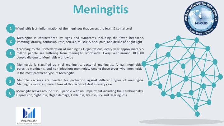
Meningitis is an inflammation of the meninges, the membranes that cover the brain and spinal cord. The infected fluid surrounding the meninges leads to the development of Meningitis. It is characterized by signs and symptoms including fever, headache, vomiting, drowsy, confusion, rash, seizure, muscle & neck pain, and dislike of bright light. Meningitis is classified as viral meningitis, bacterial meningitis, fungal meningitis, parasitic meningitis, and non-infectious meningitis.
“Defeat Meningitis”
Facts about Meningitis
According to the Confederation of Meningitis Organizations, every year approximately 5 million people are suffering from meningitis worldwide. Every year around 300,000 people die due to Meningitis worldwide.
Viral meningitis is the most prevalent type of meningitis.
Multiple vaccines are needed for protection against different types of meningitis. Meningitis vaccines prevent tens of thousands of deaths every year.
Meningitis leaves around 1 in 5 people with an impairment including the cerebral palsy, depression, sight loss, organ damage, limb loss, brain injury, and hearing loss
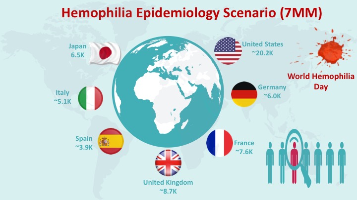
Every year, the World Hemophilia Day is celebrated on 17 April to raise awareness regarding the hemophilia and other bleeding disorders.
World Hemophilia Day is an international awareness day for hemophilia and other bleeding disorders. Hemophilia is a serious bleeding disorder in which the blood does not clot normally which results in excessive bleeding. People with severe hemophilia may bleed for no reason.
World Hemophilia Day marks the birth anniversary of Frank
Schnabel, the founder of the World Federation of Hemophilia. It is an important
day for the World Federation of Hemophila (WFH) and the bleeding disorders
community.
This year the World Hemophilia Day is celebrated in a way that is sensitive to the risks of the current global novel coronavirus (COVID-19) pandemic. This year all the activities of this day will be observed virtually to the coronavirus spread.